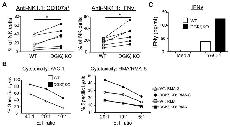
Figure 2. DGKζ KO LAK cells display increased cytotoxicity and cytokine production upon interaction with tumor cells.
A) WT or DGKζ KO LAK cells were stimulated with plate-bound anti-NK1.1 antibodies. The proportion of NK cells (CD4−CD8−NK1.1+DX5+) labeled with anti-CD107a (left) and intracellular anti-IFNγ antibody (right) is shown. Data from 6 independent experiments is shown (*P<0.05 by paired t-test). N=6 B) WT or DGKζ KO LAK cells were co-cultured with YAC-1, RMA, or RMA-S cells at the indicated E:T ratios and % specific lysis was determined 4–6 hours later. C) WT or DGKζ KO LAK cells were plated with or without YAC-1 cells at a 1:1 ratio for 24 hours. IFNγ content in the cell-free supernatants was determined by ELISA. One representative of N=3 independent experiments is shown for B and C.

