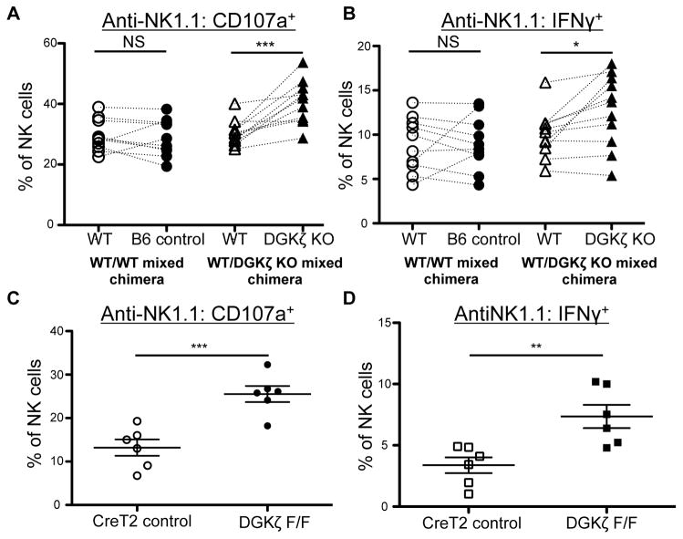
Figure 4. DGKζ KO NK cells are hyperresponsive in a cell-intrinsic and developmentally independent manner.
A) Splenocytes from WT/WT control and WT/DGKζ KO mixed BM chimeras were stimulated with plate-bound anti-NK1.1 antibody. The proportion of NK cells (CD3−DX5+NKp46+) labeled with anti-CD107a and B) intracellular anti-IFNγ antibody is shown. Cells derived from either the B6 control BM or DGKζ KO bone marrow were paired with cells that were of WT competitor BM origin within the same mouse. N=11. C) Splenocytes from Tamoxifen-treated DGKζF/F Rosa26-STOP-Flox-YFP ERCreT2 or Rosa26-STOP-Flox-YFP ERCreT2 control mice were stimulated with plate-bound anti-NK1.1 antibody. The proportion of NK cells (CD3−DX5+NKp46+YFP+ lymphocytes) labeled with anti-CD107a and D) intracellular anti-IFNγ antibody is shown. N=6. *, ** and *** represent a statistical significance by paired t-test (A and B) or Student’s t-test (C and D) of p<0.05, p<0.01 and p<0.001 respectively. NS= not significant. All data shown in this figure is compiled from at least 3 independent experiments.

