Figure 1.
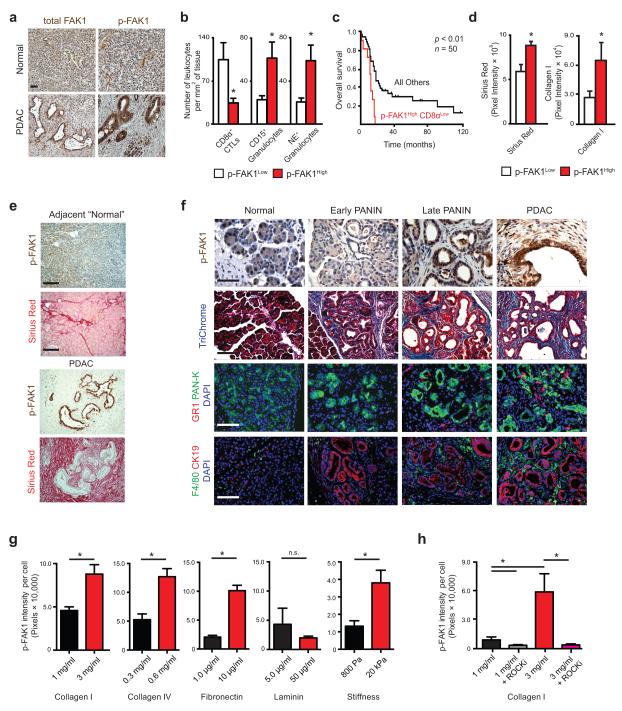
FAK1 is hyperactivated in PDAC. (a) Representative Immunohistochemistry of total and phosphorylated FAK1/FAK (p-FAK1/FAK, Tyr397) in human normal pancreas and PDAC tissues; scale bar, 100 μm. (b) Immunohistochemistry analysis of CD8α+ CTLs, CD15+ and neutrophil elastase+ (NE+) granulocyte numbers in human PDAC patient tissues subdivided into p-FAK1High (n = 33) and p-FAK1Low (n = 23) by mean p-FAK1 expression. (c) Kaplan-Meier survival analysis of PDAC patients stratified by mean p-FAK1 and CD8α+ CTL values (n = 50), with p-FAK1High CD8αLow group displayed vs. all other groups. (d) Quantification of Sirius Red staining (total collagen) or collagen I in human PDAC tumor tissues subdivided into p-FAK1High and p-FAK1Low by mean p-FAK1 expression. (e) Representative Immunohistochemistry for p-FAK1 and staining for Sirius Red in human PDAC and adjacent “normal” tissue; scale bar, 400 μm. (f) Representative immunohistochemistry for p-FAK1, Trichrome (total collagen), GR1+ granulocytes and F4/80+ TAMs in normal pancreatic tissue, early PANIN, late PANIN and PDAC tumor from KPC mice. Cytokeratin 19 (“CK19”) and Pan-Keratin (“PAN-K”) mark pancreatic epithelial cells. Scale bars: p-FAK1, 100 μm; Trichrome, GR-1 and F4/80, 200 μm. (g) Immunofluorescence analysis of p-FAK1 expression in KP cells cultured on collagen I gel, collagen IV-coated plates, fibronectin (FN1)-coated or laminin-coated polyacrylamide gels and FN1-coated compliant (800 Pa) / rigid (20 kPa) polyacrylamide gels. (h) Immunofluorescence analysis of p-FAK1 expression in KP cells cultured on collagen I gel and treated with vehicle or ROCKi (Y-27632). Error bars, mean ± s.e.m; * indicates P < 0.05 by unpaired two-sided Student’s t-test (b,d and g), log-rank test (c) or one-way ANOVA with Tukey’s method for multiple comparisons (h).

