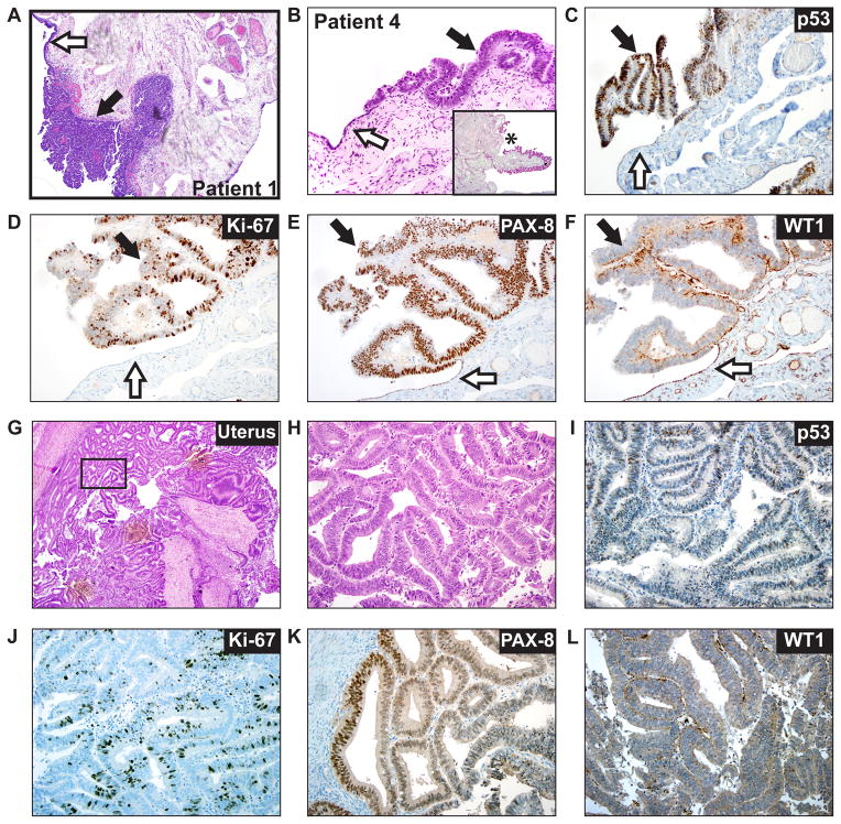
Figure 1. Intraepithelial tubal metastasis mimicking a STIC in a patient with uterine endometrial carcinoma.
A. Low power (40X) photomicrograph of a hematoxylin and eosin (H&E) stained slide from the fimbriated end of the distal fallopian tube of patient 1. The black arrow indicates the presence of STIC (noninvasive into the underlying stroma) and white arrow identifies normal adjacent tubal epithelium. B. High power (400x) photomicrograph of a H&E stained slide from the fimbriated end of the distal fallopian tube of patient 4. Inset panel shows low power (40x) micrograph of same specimen with asterisk marking area of magnification. The black arrow identifies area of significant cytologic atypia, morphologically consistent with a tubal intraepithelial carcinoma (TIC). The white arrow indicates adjacent normal tubal epithelium. C. Immunohistochemical staining (IHC) for p53 in the TIC with clonal type overexpression (black arrow). The white arrow indicates a wild type p53 staining pattern. D. IHC for Ki-67 in the TIC (black arrow) shows a mitotic index of approximately 40%. The white arrow shows low staining in the normal epithelium. E. IHC for PAX8 in the TIC (black arrow) shows strong nuclear reactivity. White arrow indicates normal epithelium. F. IHC for WT1 in the TIC (black arrow) shows absent nuclear expression. White arrow indicates WT expression in normal epithelium. G. Low power (40x) photomicrograph of an H&E stained slide from patient 4’s primary UEC. H. High power (400x) photomicrograph of the area indicated by the rectangle in panel G. I. IHC for p53 in the primary UEC shows a wild type staining pattern. J. IHC for Ki-67 in the primary UEC shows a mitotic index of approximately 20%. K. IHC for PAX-8 in the primary UEC shows patchy nuclear positivity. L. IHC for WT1 in the primary UEC shows absent nuclear expression. Next generation sequencing detected the presence of a TP53 mutation exclusively in the tubal lesion from patient 4, consistent with IHC results (see C vs. I), supporting the tubal lesion from patient 4 as a mucosal micrometastasis/implant from the patient’s primary UEC, rather than a STIC.

