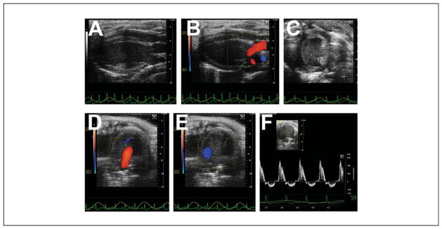
Figure 2.
Heart images from different planes. (A) Parasternal long axis view. The left ventricular apex is to our left, with the mitral valve, aortic valve, and aortic root shown. Note how the heart lies “flat” and is orthogonal to the transducer, and the normal left ventricle is shaped like a prolate ellipsoid (“bullet shaped”). (B) Parasternal long axis view, with superimposed color Doppler flow map, showing mitral inflow and aortic outflow. (C) Parasternal short axis view. The normal left ventricle is round, and systolic function is normally determined at the level of the papillary muscles. The right ventricle is often poorly seen. (D) Apical 4-chamber view, with superimposed color Doppler flow map showing mitral inflow (red jet). (E) Apical 4-chamber view, with superimposed color Doppler flow map showing aortic outflow (blue jet). (F) Pulsed-wave Doppler signal of mitral inflow plus off-angle aortic outflow.

