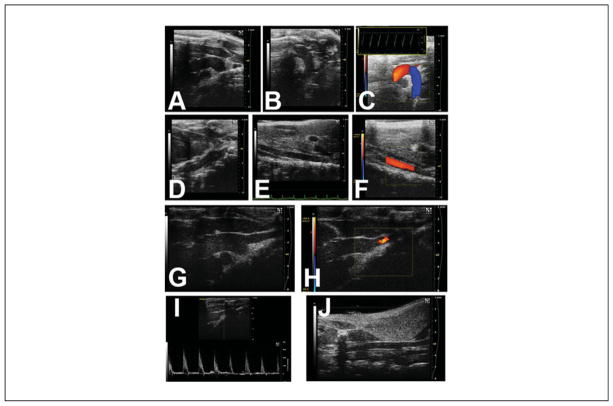
Figure 8.
Imaging of the aorta (may require multiple imaging planes) and its branches. (A) Ascending aorta and very proximal aortic arch. (B) Aortic arch and very proximal thoracic descending aorta. (C) Color Doppler flow map of aortic arch (inset: pulsed-wave Doppler signal of the distal aortic arch). (D) Thoracic descending aorta, behind the heart. (E) Abdominal aorta, near the level of the kidneys. (F) Abdominal aorta with superimposed color Doppler flow map. The brachiocephalic branches off the aortic arch may also be imaged. (G) Distal ascending aorta, with innominate artery takeoff. (H) Same as (G), but the superimposed color Doppler flow map showing normal flow into the innominate artery. (I) Pulsed-wave Doppler signal of the proximal innominate artery. (J) Right common carotid artery, further up into the neck.

