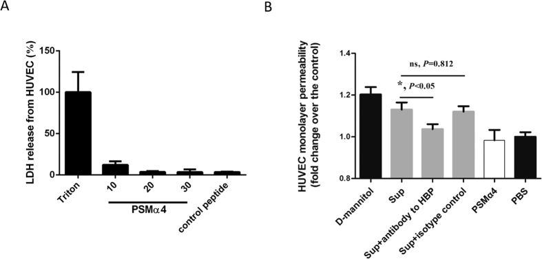
Figure 4. PSMα4-induced HBP release increases HVUEC monolayer permeability.
(A) PSMα4 did not cause cytotoxicity in HUVECs. HUVECs (104/well) were seeded in 24-well plates and incubated for 24 h. The HUVECs were then incubated with PSMα4 at 10, 20, or 30 μg/mL at 37 °C for 30 min. The supernatant was collected and cytotoxicity was analyzed by LDH assay. (B) PSMα4-induced HBP release increased HVUEC monolayer permeability. The HUVEC monolayer on the transwell insert chamber was incubated with d-mannitol (1.4 mM), culture supernatant of whole blood treated with PSMα4 (10 μg/mL), the supernatant + functional blocking antibody against HBP, the supernatant + the isotype control IgG (50 μg/mL), PSMα4 (10 μg/mL), or PBS at 37 °C for 30 min. Lucifer yellow was also added to the incubation media. The fluorescence intensity in the lower chamber of the transwells was determined. d-mannitol and PBS were used as the positive and negative control, respectively.

