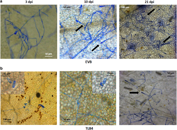
Figure 2.
Germination of E. necator conidia in the control line EVB (a) and in the resistant transgenic line TLB4 (b). Microscopy images of infected leaves were taken at 3, 10 and 21 days post inoculation (d.p.i.) with powdery mildew. Insert at high magnification highlights the germination of an E. necator conidia at 3 and 10 d.p.i. The arrows indicate the conidiophores.

