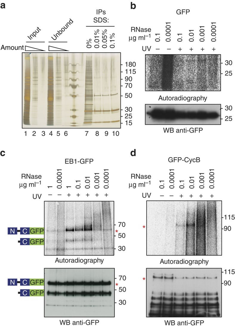
Figure 3. Validation of CycB and EB1 as RNA-binding proteins.
(a) Optimization of GFP immunoprecipitation. Protein profiles of total embryo lysates, IPs of free GFP and unbound fractions are visualized on a silver stained polyacrylamide gel. Lanes 1–3: serial dilution of total embryo lysate expressing free GFP; lanes 4–6: serial dilution of unbound fractions after GFP-IP; lanes 7–10: GFP immunoprecipitates washed with buffers containing increasing concentrations (none; 0.01%; 0.05%; 0.1%) of SDS. (b–d) Lysates containing free GFP, EB1-GFP and GFP-CycB were treated with various concentrations of RNase A. GFP proteins were IPed from noCL and CL embryos, radioactively labelled, separated on polyacrylamide gels and blotted on PVDF membranes. Upper panels: autoradiography of membranes containing IPs of GFP (b), EB1-GFP (c) and GFP-CycB (d) labelled with γ-[32P]-ATP by T4 polynucleotide kinase (see Methods section, PNK assay). Lower panels: visualization of GFP proteins by WB with an anti-GFP antibody.

