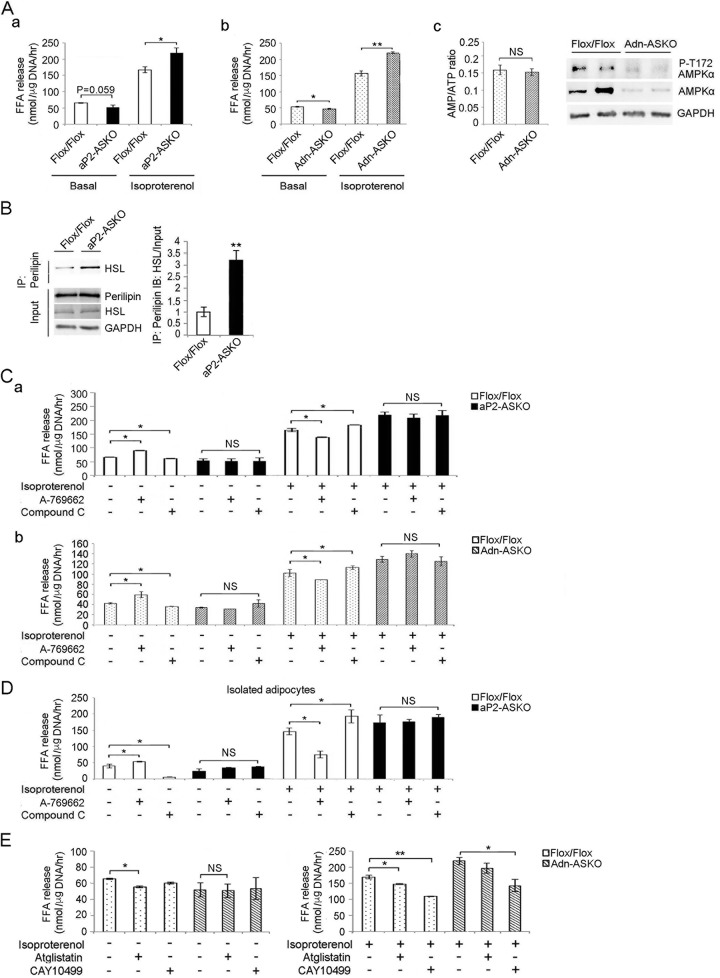
FIG 4.
AMPK induces basal lipolysis and inhibits stimulated lipolysis. (A) FFA release from WAT of aP2-ASKO (a) and Adn-ASKO (b) mice under basal and isoproterenol-stimulated conditions. Cellular AMP/ATP ratios in WAT of Flox/Flox and Adn-ASKO mice under basal conditions (c, left) and immunoblotting for AMPKα and AMPKα phosphorylated at T172 (c, right). (B) Immunoblotting with HSL antibody after immunoprecipitation (IP) of WAT lysates with perilipin antibody. (C) FFA release from WAT of aP2-ASKO (a) and Adn-ASKO (b) mice upon treatment with 2 mM A-769662 or 50 μM compound C under basal or stimulated conditions (n = 5). (D) FFA release from isolated adipocytes of aP2-ASKO mice upon treatment with 2 mM A-769662 or 50 μM compound C under basal and stimulated conditions (n = 4). (E) FFA release from Adn-ASKO mouse WAT explants upon treatment with 50 μM atglistatin or 10 μM CAY10499 under basal and isoproterenol-stimulated conditions. Data are expressed as means ± SEM. *, P < 0.05; **, P < 0.01; NS, no significant change. Experiments were repeated twice, and representative data are shown.

