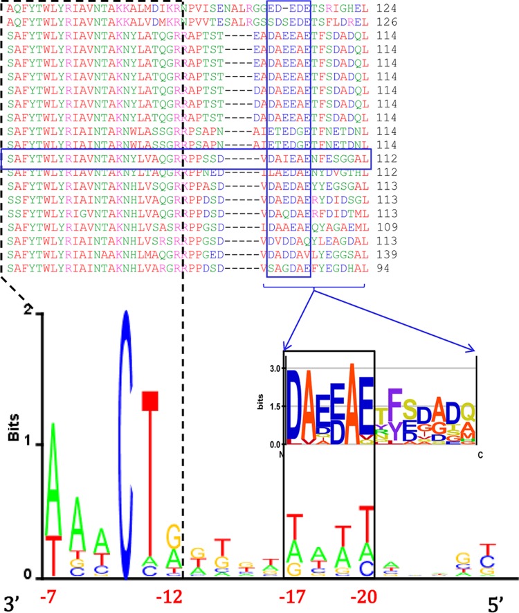
FIG 6.
Putative σE protein-DNA spacer interactions. (Top) Multiple alignment of the protein sequence of σE (boxed in blue) and those of selected ECF02 members (the corresponding GI numbers are provided in Table S1 in the supplemental material), showing the C-terminal part of the σ2 domain and the flanking sequence C-terminal of domain σ2. The conserved protein motif identified is boxed in blue. Below the alignment is a sequence logo presenting the conserved protein motif and the (unconserved) flanking sequence C-terminal of that motif. (Bottom) The −10 element of the promoter, together with the spacer sequence, is presented in the form of a 3′-to-5′-oriented sequence logo (coordinates are indicated relative to the transcription start site). The putative interaction between the conserved protein motif and the conserved promoter spacer motif is indicated by outlining (solid black lines), as is the −10 element–σ2 domain interaction (dashed black lines).

