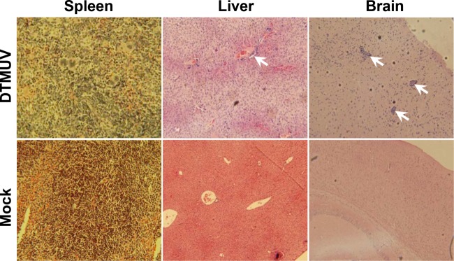
FIG 7.
Histopathological assays of major target organs in DTMUV-infected IFN-α/βR−/− mice. Four-week-old female IFN-α/βR−/− mice were inoculated by the i.p. route with 105 PFU of DTMUV or PBS as a control. At 6 days p.i., the brains, livers, and spleens of mice were collected, fixed with perfusion fixative (4% formaldehyde) for 48 h, and processed according to standard histological methods. All sections from each tissue were stained with H&E. Arrows, perivascular and parenchymal mononuclear inflammatory cell infiltration and neuronal degeneration.

