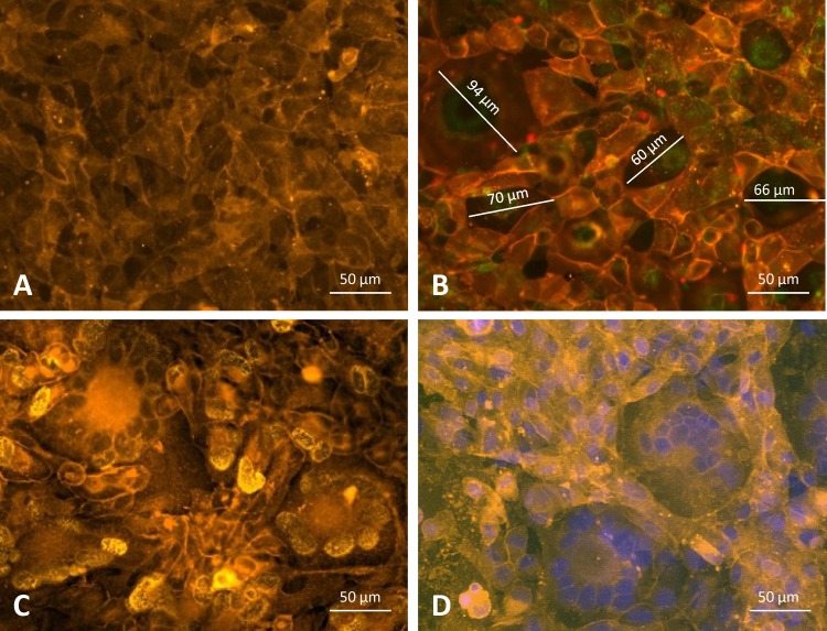
FIG 2.
Monolayers of ARPE-19 epithelial cell cultures. (A) Uninfected ARPE-19 cells stained with the yellow-orange fluorescent dye PKH-26 and interacting with plasma membrane lipids. (B) VR1814-infected cell culture stained with PKH-26, showing large syncytia (a single diameter is reported for each syncytium) as well as single membrane-limited cells. (C) VR1814-infected cell culture stained with PKH-26 and an HCMV p72 MAb, showing true SF and limiting membranes. (D) VR1814-infected cell culture stained with PKH-26 and DAPI.

