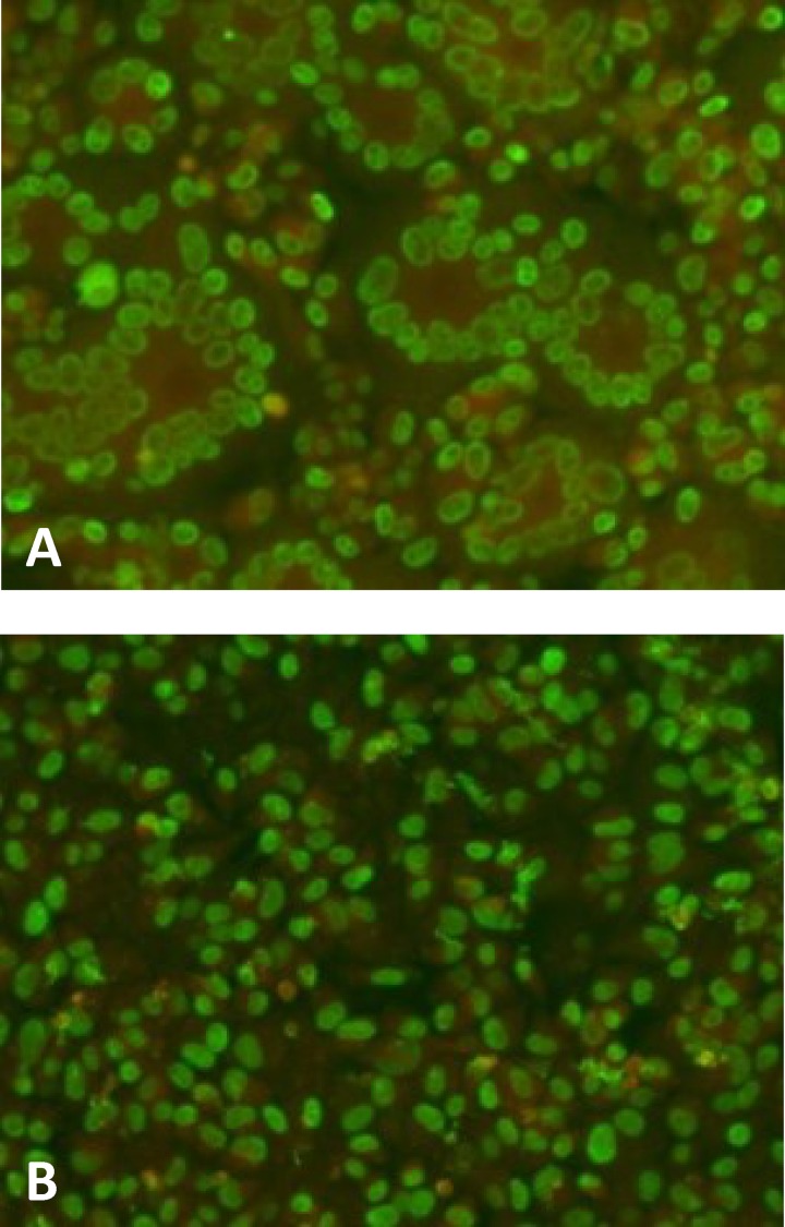
FIG 3.
Immunofluorescence staining of VR1814-infected ARPE-19 epithelial cells at 96 h p.i. HCMV fluorescent nuclei (p72) are shown in green, while Evans blue counterstaining is shown in red. (A) Monolayer of ARPE-19 cells. Several syncytia are seen throughout the cell monolayer. (B) One hundred percent SF blocking by a human MAb (4I22; 10 μg/ml) directed to pUL130-131 of the pentamer.

