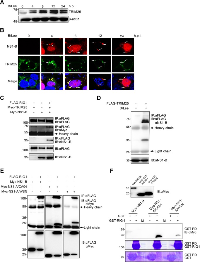
FIG 5.
NS1-B is involved in a RIG-I/TRIM25/NS1-B ternary complex by interacting with TRIM25. (A) A549 cells were infected at an MOI of 0.5 with B/Lee. At the indicated time points, the cell lysates were harvested and quantified with a BCA protein assay kit. The amount of TRIM25 was tested by immunoblotting with anti-TRIM25 antibody. (B) 293T cells were transfected with FLAG-TRIM25 for 24 h, followed by infection with B/Lee at an MOI of 0.5 for the indicated times. The localization of FLAG-TRIM25 (green) and NS1-B (red) was determined using fluorescence microscopy with an anti-FLAG monoclonal antibody and anti-NS1-B polyclonal antibody. The nuclei were stained with DAPI. White arrows indicate the granule-like aggregates. (C) 293T cells were transfected with FLAG-RIG-I, together with Myc-TRIM25, Myc-NS1-B, or both, for 36 h. Whole-cell lysates of all the samples were subjected to immunoprecipitation with anti-FLAG, followed by IB with anti-FLAG, anti-Myc, and anti-NS1-B antibodies. (D) 293T cells were transfected with FLAG-TRIM25 for 24 h, followed by infection with B/Lee at an MOI of 0.5 for 4 h. The cell lysates were harvested and subjected to immunoprecipitation with an anti-FLAG antibody, followed by IB with anti-FLAG and anti-NS1-B antibodies. (E) Whole-cell lysates of 293T transfected with FLAG-RIG-I or FLAG, together with Myc-NS1-B or Myc-NS1-A (NS1-A/CA04 and NS1-A/WSN), for 24 h were subjected to immunoprecipitation with an anti-FLAG antibody, followed by IB with anti-FLAG and anti-Myc antibodies. (F) Whole-cell lysates of 293T cells were transfected with Myc-NS1-B or Myc-NS1-A (NS1-A/CA04 and NS1-A/WSN) for 48 h mixed with GST–RIG-I or GST for GST pulldowns (GST-PD), followed by IB with an anti-Myc antibody or Coomassie staining. M, protein marker.

