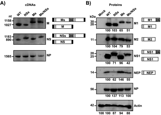
FIG 2.
Characterization of the influenza PR8 viruses harboring split sequences. MDCK cells were infected (MOI, 3) with WT PR8 virus or PR8 viruses harboring split segments (NSs, Ms, and Ms/NSs), and at 18 h postinfection, cells were harvested to evaluate RNA (A) or protein (B) expression levels. (A) cDNA synthesis and PCRs for NS, M, or NP viral segments were performed using specific primers. (B) Protein expression levels for M1, M2, NS1, NEP, and NP were evaluated using protein-specific antibodies. Actin was used as a loading control. Numbers to the left of the gels indicate the sizes (in nucleotides) of cDNA (A) and the sizes (in kilodaltons) of proteins (B). A schematic representation of the viral segments (A) or protein products (B) is provided on the right of each panel. Western blots were quantified by densitometry using the software ImageJ (v.1.46). Protein expression in WT-infected cells was considered 100% for comparison with the level of expression by the corresponding viruses harboring split segments (indicated by the numbers [in percent] below each blot in panel B).

