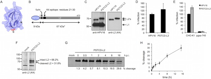
FIG 1.
Characterization of the PSTCD-L2 virus. (A) Atomic structure of the 70-residue fragment of the PSTCD domain, modeled with cartoon and mesh surface rendering. The position of the C terminus that is fused to L2 is shown in red. (B) Schematic of the PSTCD-L2 fusion, indicating the positions of the furin cleavage site, the K4 epitope, and predicted cleavage fragment sizes. The asterisk denotes the apparent molecular mass of 67 kDa; the calculated molecular mass is 49 kDa. (C) Western blot of CsCl-purified PSTCD-L2 virions, compared to wt HPV16. L1 and L2 were detected with rabbit anti-HPV16 polyclonal antibody, and L2 was detected with mouse anti-L2 K4 monoclonal antibody. (D and E) Relative infectivity of wt or PSTCD-L2 virus in HaCaT cells (D) or CHO-K1 and HSPG-deficient pgsa-745 cells (E). (F) Representative Western blot of mock- and PSTCD-L2 infected HaCaT cell lysates 24 h postinfection, stained with the K4 monoclonal antibody. Cleavage levels were measured by densitometry. Values are the means from three independent experiments. (G) Representative time course blot of PSTCD-L2 cleavage in HaCaT cells. (H) Plot of PSTCD-L2 cleavage levels from five independent time course experiments. Values are means ± standard deviations.

