
FIG 8.
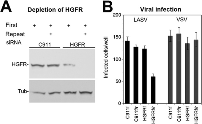
Infection of rLCMV-LASVGP in A549 cells depleted of HGFR. (A) Depletion of HGFR by RNAi. A549 cells were transfected with siRNAs specific for HGFR and the corresponding control siRNA (C911). An initial transfection (First) was performed 16 h after plating. Where indicated, a second (Repeat) transfection was performed after another 24 h. Expression of HGFR was determined 72 h after the first transfection by Western blotting. α-Tubulin (Tub) was detected as a loading control. Note the only partial depletion of HGFR observed after a single transfection with siRNA. (B) Viral infection of HGFR-depleted cells. Cells were transfected with the indicated siRNAs as described in the legend to panel A, with the first transfection only (f) and the combined first and repeat transfections (f/r) being indicated. At 72 h posttransfection, cells were infected with rLCMV-LASVGP (LASV) and rLCMV-VSVG (VSV) (200 PFU/well). Infection was detected after 16 h by IFA for LCMV NP as described in the legend to Fig. 2B. Data are means ± SDs (n = 3).