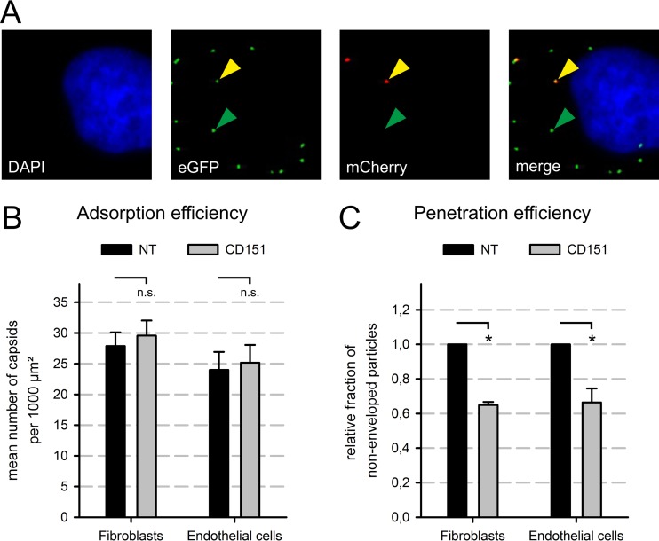
FIG 5.
CD151 depletion affected viral penetration but not adsorption. (A) Exemplary images showing fluorescence signals in a cell infected with the dual-fluorescence virus strain TB40-BACKL7-UL32eGFP-UL100mCherry. Green signals were interpreted as EGFP-labeled capsids (green arrowheads) and red signals as mCherry-labeled viral envelopes. In the merged image, particles showing both green and red signals appear yellow (yellow arrowhead). The cell nucleus was counterstained with DAPI (blue). (B and C) Fibroblasts (HFFs) and endothelial cells (HEC-LTTs) were transfected for 48 h with CD151 or NT siRNA and then inoculated with the dual-fluorescence HCMV strain TB40-BACKL7-UL32eGFP-UL100mCherry at an MOI of 20 to 80. Cells were fixed after 30 min. Fluorescence images were taken and analyzed by determining the total number of green signals (all capsids, regardless of the presence or absence of envelope) (B) and the fraction of green-only signals, without red (nonenveloped particles) (C), to determine the total number of adsorbed virus particles (B) and the fraction of successfully penetrated particles (C). In each experiment, >800 particles were counted for each condition. The penetration efficiency was standardized to the NT control level. Error bars show standard errors of the means. n.s., not significant; *, significant (P < 0.05; Student's t test).

