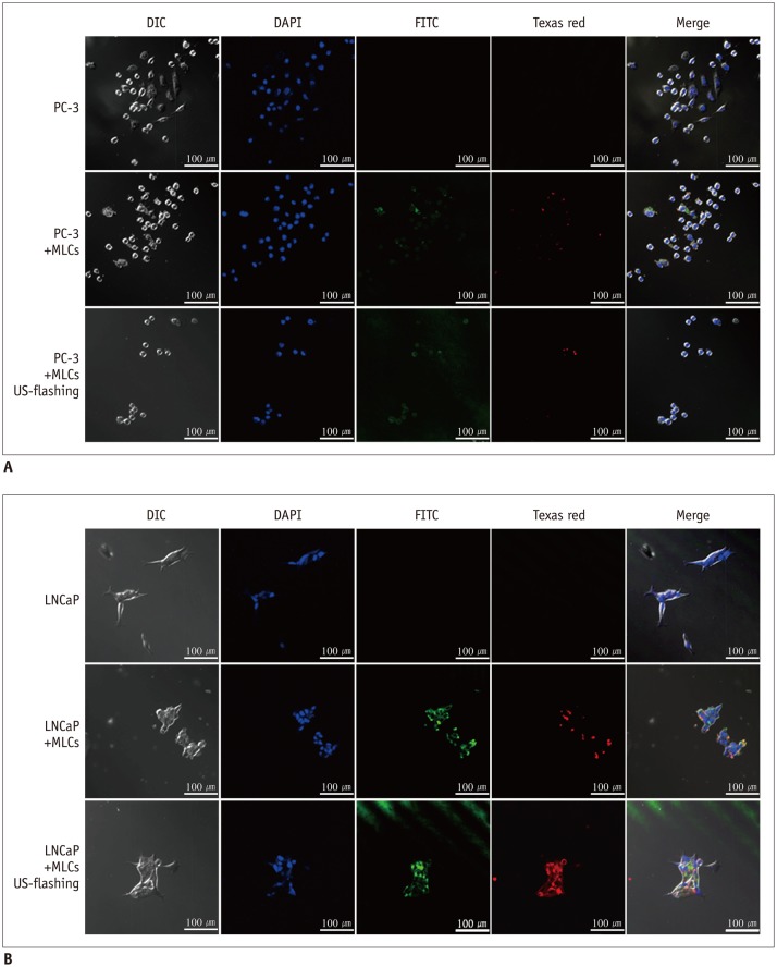
Fig. 2. Confocal laser scanning microscopy images of PC-3 cells and LNCaP cells.
A. Confocal microscopy images reveal no visible fluorescence in cells (× 400 magnification), suggesting poor uptake of MLCs into PC-3 cells. B. Green fluorescence in cells labeled by FITC and red fluorescence in cells labeled by Texas red are observed under microscopy (× 400) before and after ultrasound exposure. Observed fluorescence patterns suggest that microbubble-liposome complexes (MLCs) conjugated with anti-Her2 antibodies efficiently target LNCaP cells. Her2 = human epidermal growth factor receptor type 2

