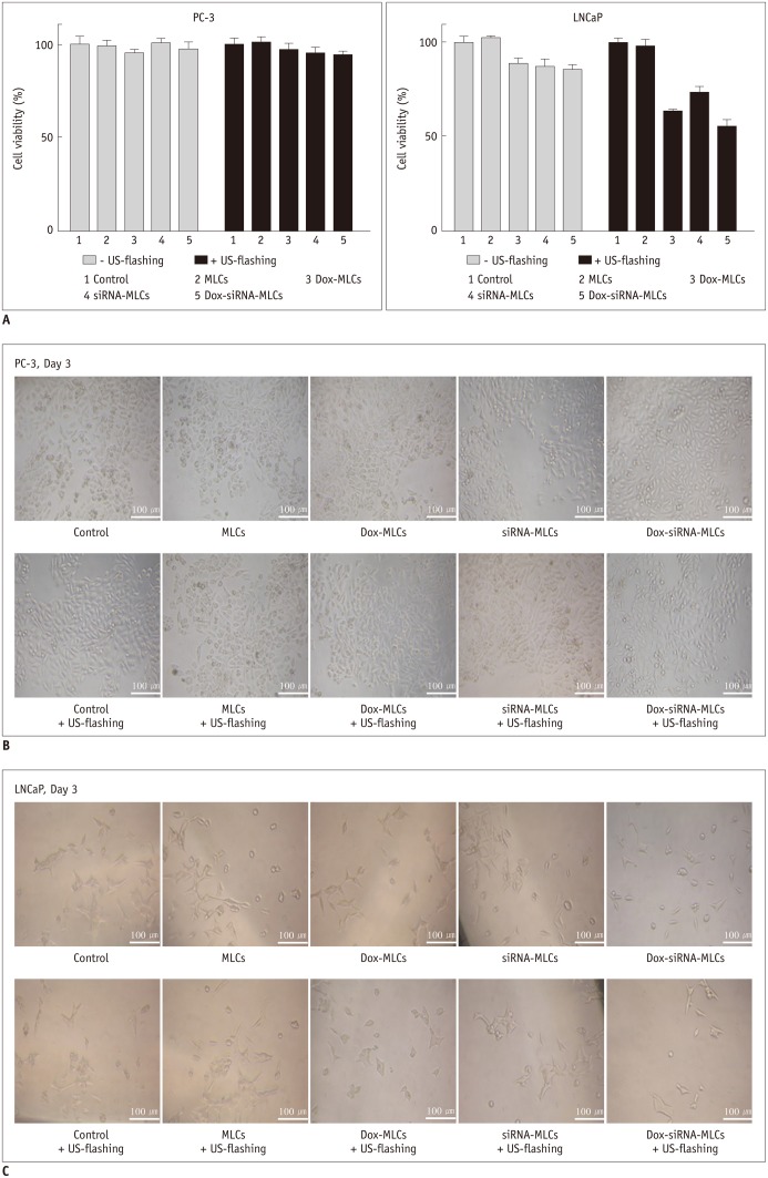
Fig. 4. Effect of therapeutic agents and ultrasound guidance on viability of PC-3 and LNCaP cells.
A. Bar graph depicting viability of PC-3 and LNCaP cells, demonstrating that viability of LNCaP cells was decreased following ultrasound exposure when treated with Dox-MLCs (from 88.0 ± 3.4% to 63.0 ± 1.8%), siRNA-MLCs (from 87.0 ± 4.1% to 73.0 ± 3.8%), and Dox-siRNA-MLCs (from 85.0 ± 2.9% to 55.0 ± 3.5%). All decreases were statistically significant (p < 0.01). Dox-MLCs = MLCs with doxorubicin, Dox-siRNA-MLCs = MLCs with siRNA and doxorubicin, MLCs = microbubble-liposome complexes, siRNA-MLCs = MLCs with siRNA B, C. MTT assays performed with PC-3 and LNCaP cells show no significant difference in viability of PC-3 cells between treatment subgroups. Conversely, cell viabilities were decreased after ultrasound exposure in subgroups of LNCaP cells. Dox-MLCs = MLCs with doxorubicin, Dox-siRNA-MLCs = MLCs with siRNA and doxorubicin, MLCs = microbubble-liposome complexes, siRNA-MLCs = MLCs with siRNA

