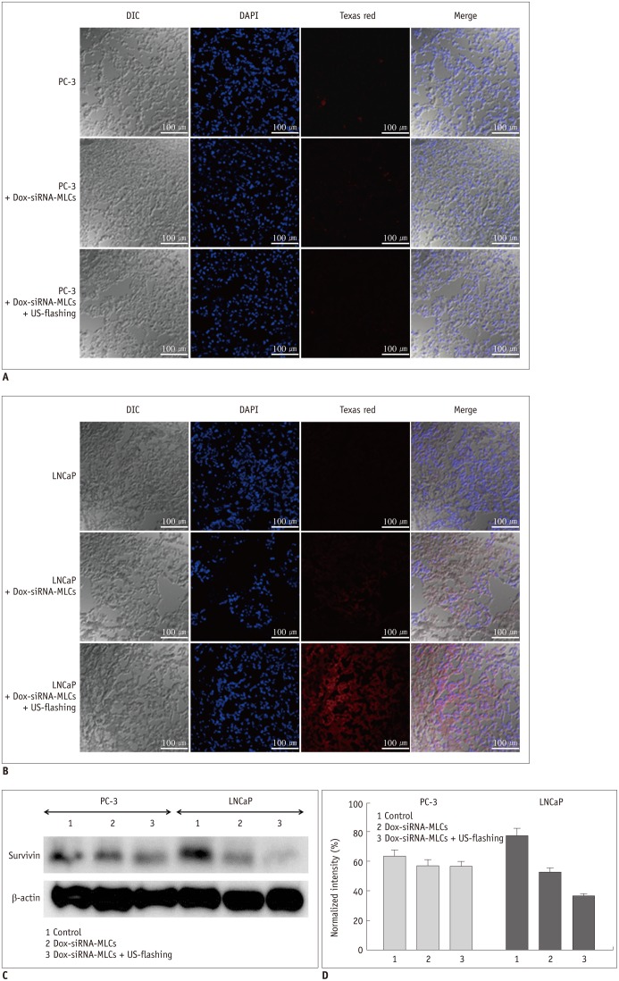
Fig. 5. Effect of treatment with microbubble-liposome complex (MLC) on PC-3 and LNCaP xenograft tumor models.
A. No fluorescence signal was observed in PC-3 tumor on confocal microscopy (× 400 magnification). Dox-siRNA-MLCs = MLCs with siRNA and doxorubicin B. Bright red fluorescence signal was observed in LNCaP tumor in confocal images (× 400), suggesting intra-tumor uptake of MLCs. It should be noted that amount of intra-tumor uptake of fluorescent MLCs after ultrasound exposure is increased after ultrasound exposure. C. Western blot analysis demonstrated reduced survivin expression in LNCaP cells treated with siRNA-loaded MLCs. Levels of expression of survivin are further decreased following ultrasound exposure. Protein expression was normalized to expression of β-actin. D. Bar graphs show mean survivin density in each treatment group. Mean survivin density is lower in treated LNCaP cells compared to control cells (group 1, 77.4 ± 4.90%; group 2, 52.7 ± 2.83%; group 3, 36.7 ± 1.34%; p = 0.027). No substantial decrease in density of survivin was observed in PC-3 cells (group 1, 63.1 ± 4.36%; group 2, 56.8 ± 4.35%; group 3, 56.6 ± 3.08%; p = 0.113). Dox-siRNA-MLCs = MLCs with siRNA and doxorubicin

