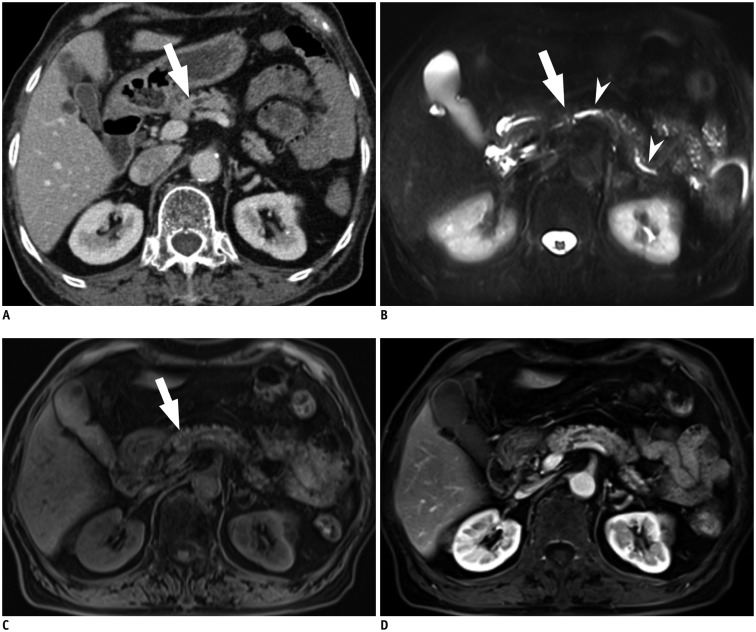
Fig. 1. 81-year-old woman with pancreatic ductal adenocarcinoma.
A. Axial, post-contrast CT image obtained during hepatic venous phase shows dilatation of main pancreatic duct with abrupt narrowing (arrow) at neck portion of pancreas. However no definite focal pancreatic mass lesion obstructing main pancreatic duct is visualized on CT. Both reviewers answered that SPL per se was not detected on CT images. However, main pancreatic ductal change led both reviewers to detection of SPL in consideration of secondary features and to correct diagnosis. B. Axial, fat-suppressed, T2-weighted image demonstrates dilatation of main pancreatic duct (arrowheads) and abrupt narrowing (arrow) without definite parenchymal lesion at neck portion of pancreas. C. Axial, fat–suppressed, T1-weighted image shows approximately 1 cm sized ill-defined, subtle hypointensity lesion (arrow) in neck portion of pancreas. D. Axial, post-contrast, T1-weighted image obtained during pancreatic phase, shows no definite mass lesion in pancreas. In addition, diffusion-weighted image did not show definite focal lesion in pancreas (not shown). Both reviewers reported SPL per se as detectable on MR image with poor lesion conspicuity graded as 1, and their specific diagnosis was pancreatic ductal adenocarcinoma. SPL = solid pancreatic lesion

