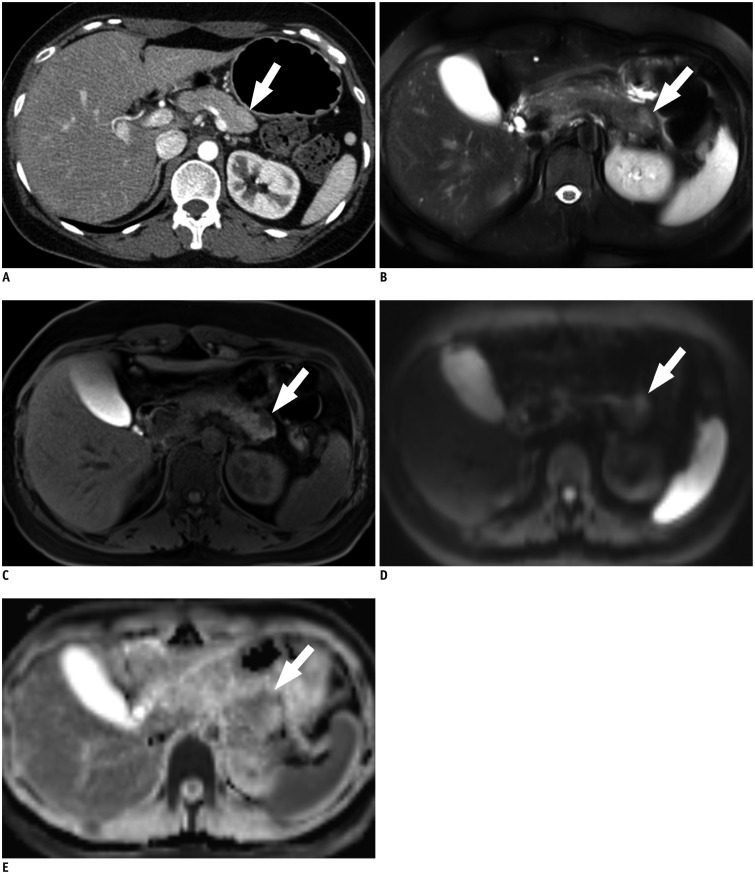
Fig. 3. 40-year-old woman with pancreatic neuroendocrine tumor, grade 1.
A. Axial, post-contrast CT image obtained during pancreatic phase shows subtle, slightly high attenuated lesion (arrow) in tail portion of pancreas. On CT, both reviewers were not able to detect "SPL in consideration of secondary features" as well as "SPL per se". B. Axial, fat-suppressed, T2-weighted image demonstrates approximately 1.5 cm sized hyperintense lesion (arrow) at pancreatic tail. C. On axial, fat–suppressed, non-enhanced T1-weighted image, lesion (arrow) shows marked hypointensity compared to that of pancreas parenchyma. D, E. Axial, diffusion-weighted image with b value of 800 sec/mm2 (D) and apparent diffusion constant map (E) also demonstrate hyperintense lesion (arrows) with diffusion restriction. On MRI, both reviewers were able to detect SPL per se and made correct specific diagnosis. Lesion conspicuity was excellent for reviewer 1, and good for reviewer 2. SPL = solid pancreatic lesion

