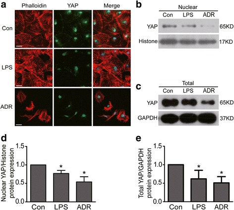
Fig 4.

YAP expression was reduced in LPS or ADR-injured podocytes. a Confocal images of podocytes showing expression of YAP (green), Phalloidin-stained stress fiber (red) and DAPI-stained nuclei (blue). Compared to controls, nuclear and cytoplasmic YAP and stress fiber were reduced in LPS or ADR-injured podocytes. Scale bar = 50 μm. b & d WB results show that nuclear YAP protein expression was reduced in both LPS and ADR treated podocytes. c & e WB results show that total YAP protein expression was reduced in both LPS and ADR treated podocytes. All results were expressed as fold YAP of LPS or ADR group in relation to controls normalized to Histone or GAPDH. Data were from at least three independent experiments. * P < 0.05 vs controls
