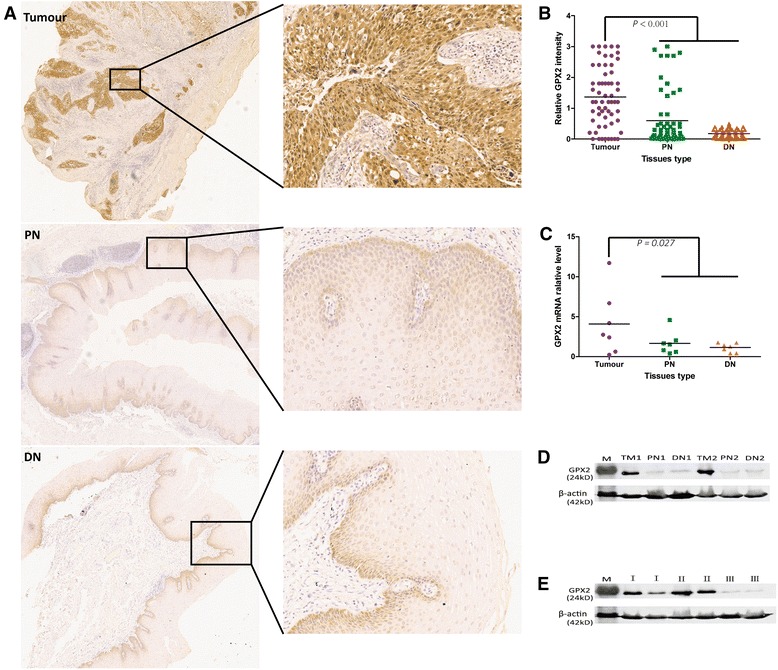
Fig. 2.

GPX2 overexpression in tumour tissues compared with non-tumour tissues including PN and DN tissues. a Representative IHC images of GPX2 staining in tumour, PN and DN tissues. b The immunochemistry analysis of the relative expression of GPX2 in tumour, PN and DN tissues according to staining intensity. There is a statistically significant difference in GPX2 protein expression of tumour tissues compared with compared with non-tumour tissues including PN and DN tissues (P < 0.001) studied by the independent-samples test. c The relative level of GPX2 mRNA expression in tumour, PN and DN tissues. There is statistically significant difference in GPX2 mRNA expression of tumour tissues compared with compared with non-tumour tissues including PN and DN tissues (P = 0.027) studied by the independent-samples test. d Western blot analysis of GPX2 protein in tumour, PN and DN tissues from two patients within ESCC. Obviously, GPX2 protein was overexpressed in tumor tissues compared with PN and DN tissues. e Western blot analysis of GPX2 protein in ESCCI, ESCCII and ESCCIII tissues from six patients with ESCC. Obviously, the expression of GPX2 protein was down-regulated in ESCCIII tissues compared with ESCCI and ESCCII tissues. M: protein marker; TM: tumour;I: ESCCI; II: ESCCII; III: ESCCIII
