Fig. 3.

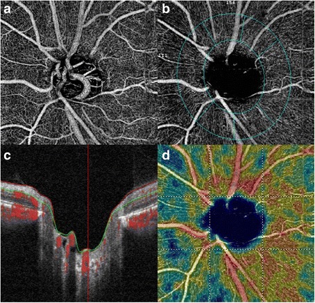
Optical coherence tomography angiography of the left optic disc, suggesting early glaucomatous optic nerve damage (a). The in-built software allows analysis of the peripapillary vascular flow and nerve fibre layer (b) suggesting neuroretinal rim thinning(c) and reduction in peripapillary retinal perfusion (d)
