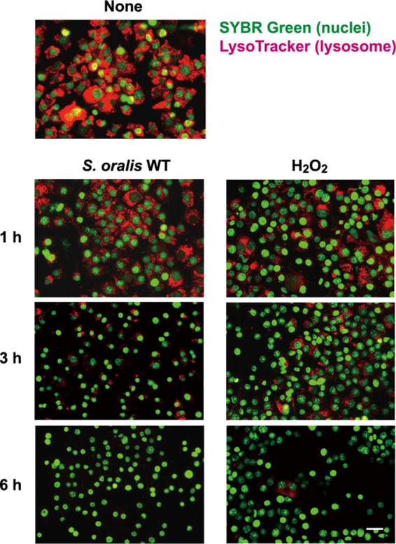
FIG 3.

S. oralis and H2O2 mediate lysosomal damage. RAW 264 cells were exposed to the S. oralis WT or H2O2 for 3 h. Then, the cells were cultured for an additional 3 h (total, 6 h) in fresh medium containing antibiotics. At 1, 3, and 6 h after exposure, the cells were stained with LysoTracker red and SYBR green II. Lysosomal deacidification was monitored using LysoTracker red (red), which accumulates in acidic organelles. Bar = 20 μm.
