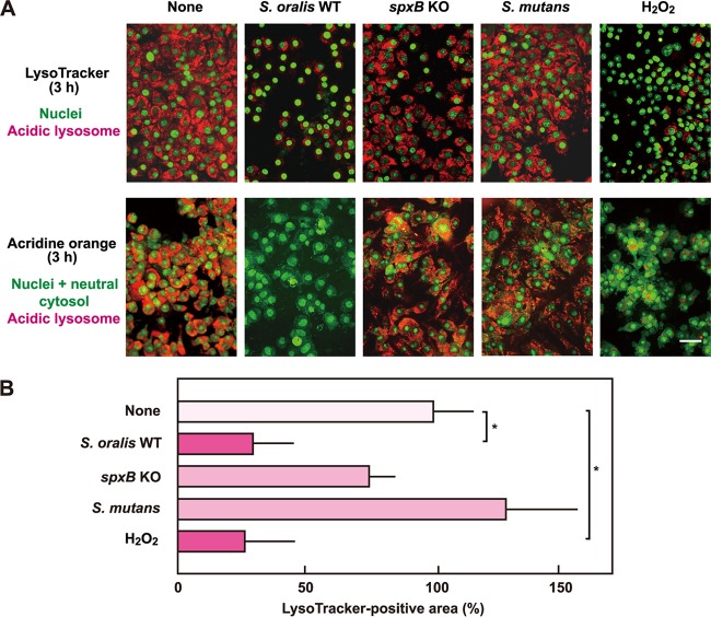
FIG 4.
The H2O2 produced by S. oralis induces lysosomal deacidification. (A) RAW 264 cells were exposed to the S. oralis WT or spxB KO mutant, S. mutans MT8148, or H2O2 for 3 h. (Top) The cells were stained with LysoTracker red and SYBR green II or acridine orange. LysoTracker red accumulates in acidic organelles. (Bottom) The acidic environment in lysosomes was also monitored by acridine orange staining. Acridine orange emits red fluorescence in acidic environments, and thus, the disappearance of red fluorescence reflects lysosome deacidification. Bar = 20 μm. (B) Measurements of the LysoTracker-stained fluorescent areas were conducted using ImageJ software. The average fluorescence area of control cells (None) was set to 100%. The results are shown as the mean ± SD for four samples. *, P < 0.05 compared with the untreated control (None).

