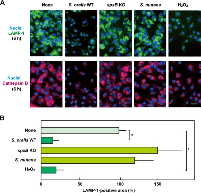
FIG 6.
The H2O2 produced by S. oralis induces lysosomal destruction. (A) RAW 264 cells were exposed to the S. oralis WT or spxB KO mutant, S. mutans MT8148, or H2O2 for 3 h. The cells were then washed and cultured for an additional 3 h in fresh medium containing antibiotics. (Top) LAMP-1 and DNA were labeled with Alexa Fluor 488-conjugated anti-LAMP-1 monoclonal antibody and DAPI, respectively. (Bottom) Cathepsin B was also labeled with Alexa Fluor 594-conjugated anti-goat IgG. Bar = 10 μm. (B) Measurements of the LAMP-1-positive areas were conducted using ImageJ software. The average fluorescence area for untreated control cells (None) was set to 100%. The results are shown as the mean ± SD for four samples. *, P < 0.05 compared with the control (None).

