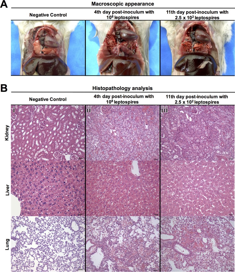
FIG 2.
Pathology of hamsters infected intraperitoneally with strain Fiocruz L1-130. (A) Representative photographs of gross examination of a negative-control hamster and hamsters infected with 108 leptospires (high dose) or 2.5 × 10² leptospires (low dose) at days 4 and 11 postchallenge, respectively. Infected hamsters had evident localized hemorrhage of the lungs. (B) Histopathology analysis showing representative photomicrographs of hematoxylin- and eosin-stained sections of kidney, liver, and lung of a negative-control animal (i) and animals infected with a high dose (ii) or a low dose (iii) of leptospires at days 4 and 11 postchallenge, respectively. The histopathology photomicrographs were taken at a magnification of ×400. Infected animals had similar histopathological features, with kidneys showing mild hyaline degeneration and hemorrhage, livers with mild loss of parenchymal architecture and steatosis, and lungs with hemorrhage. Bars, 100 μm for tissues of negative-control animal and 50 μm for tissues of animals infected with a high or a low dose, with exception of liver, which has a scale bar of 25 μm.

