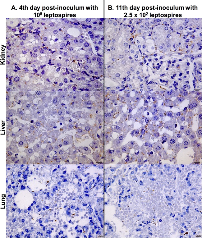
FIG 3.
Representative photomicrographs of immunohistochemically stained sections of kidney, liver, and lung from hamsters infected intraperitoneally with strain Fiocruz L1-130. Shown are stained tissues of an animal infected with 108 leptospires (high dose) at day 4 postchallenge (A) and an animal infected with 2.5 × 10² leptospires at day 11 postchallenge (B). Detection was performed with monoclonal antiserum specific for LipL32. The photomicrographs were taken at a magnification of ×1,000 and show whole leptospires and degraded cells in kidney tubules, interstitium of the liver, and alveolar septa of the lung. Bar, 20 μm.

