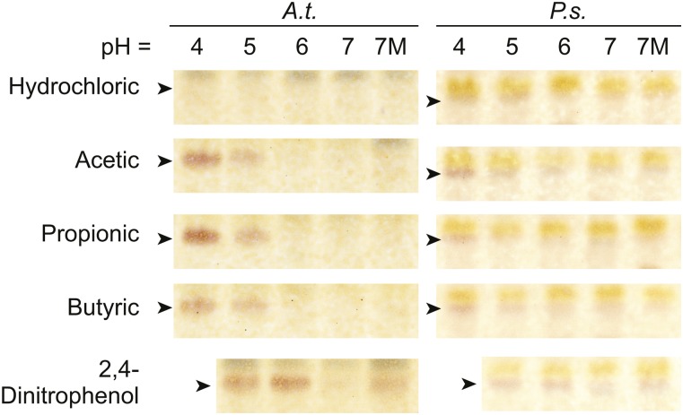
Figure 2.
pH and magnesium changes activate SFR in whole tissues. Thin-layer chromatograms show the separation of lipids from extracts of Arabidopsis (A.t.) shoots or pea (P.s.) leaves floated on 20 mm of the acid indicted at left adjusted to the pH indicated at top with dipotassium phosphate for 1 h. 7M indicates pH 7 with an additional 20 mm MgCl2. TGDG is indicated by arrowheads. Images shown are representative of three separate plant growth trials.

