Figure 6.
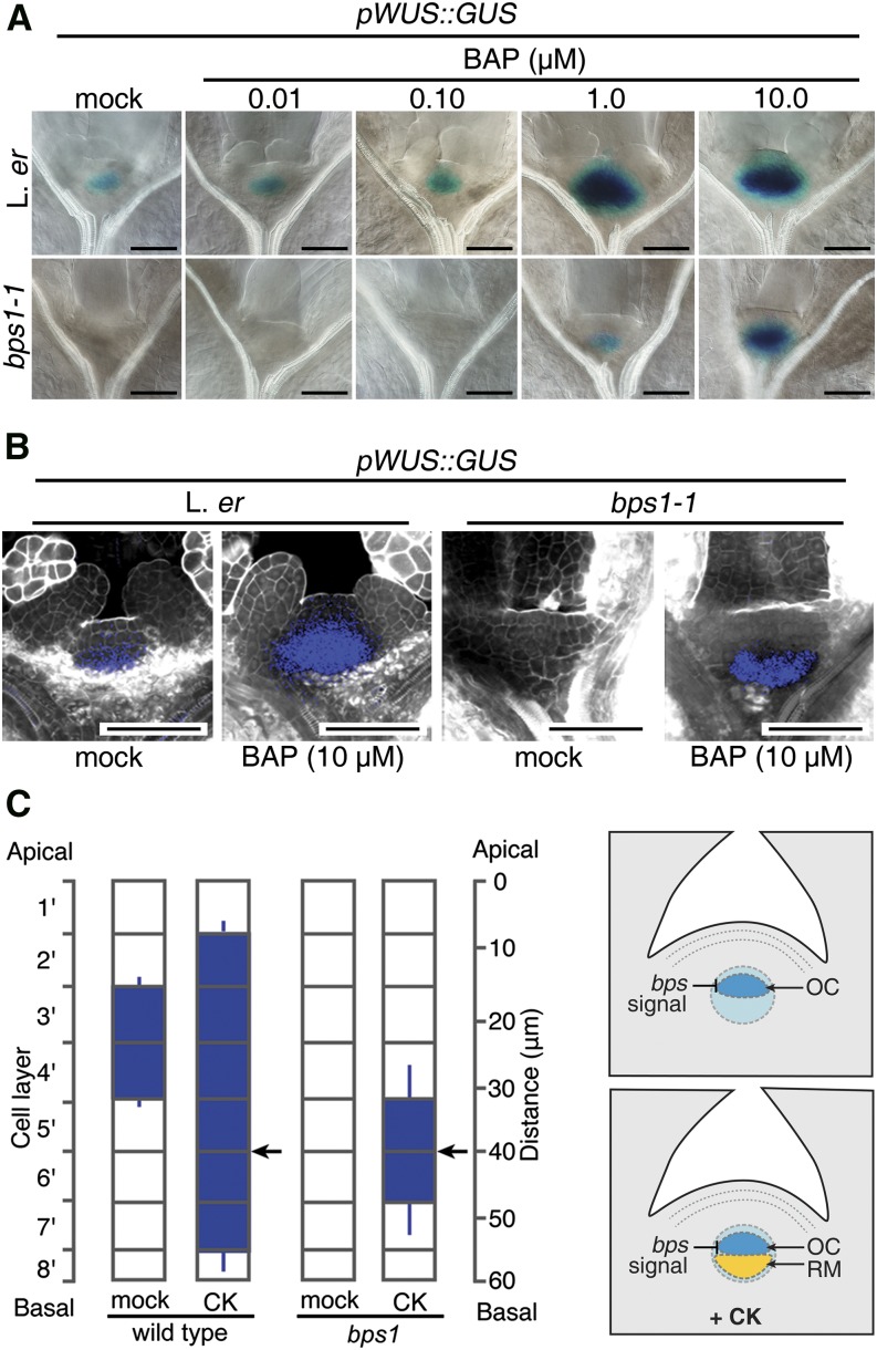
CK treatment restores WUS expression in the bps1 rib meristem. A, Shoot expression of the pWUS::GUS in 5-d wild-type (Ler) and bps1-1, mock-treated or transferred to 0.01 to 10 µM CK (BAP) for 24 h. Bars = 50 µm. B, Confocal images of pWUS::GUS expression (4 dpi) in mock-treated or in seedlings transferred to 10 µM BAP for 24 h. C, Quantitative analysis of CK-treated pWUS::GUS expression domain positions; scale on the left indicates cell layers, and scale on the right indicates distance from the apex (n = 20). Arrows point to strongest GUS staining (error bar, sd). Cartoon to the right depicts the two domain model for WUS expression.

