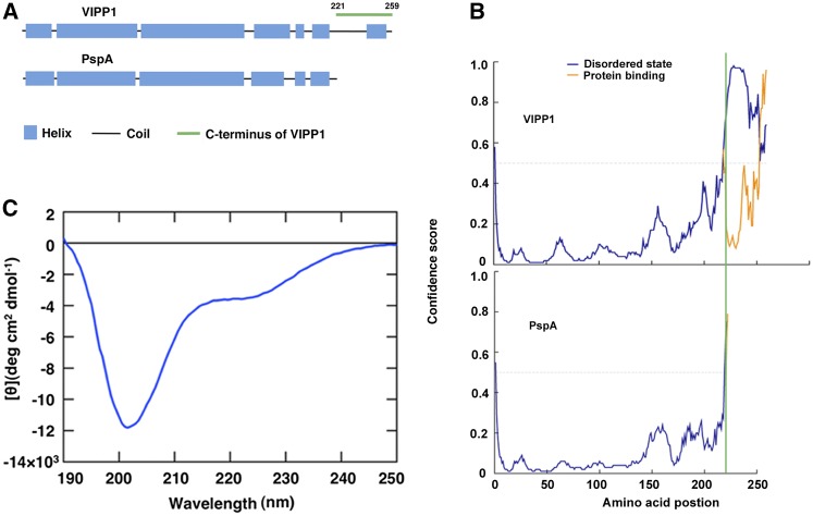
Figure 1.
Intrinsically disordered property of Vc. A, Comparison of secondary structures between VIPP1 and PspA predicted by PSIPRED. Blue and black bars, respectively, represent the α-helix and the random coil. A peptide corresponding to Vc (from 221 to 259 amino acids) is marked by a green line. B, Probability plot of IDR in VIPP1 (top) and PspA (bottom) using PSIPRED. The disordered stretch corresponding to Vc is to the right side of the green line. C, CD spectra of 190 to 250 nm at room temperature.

