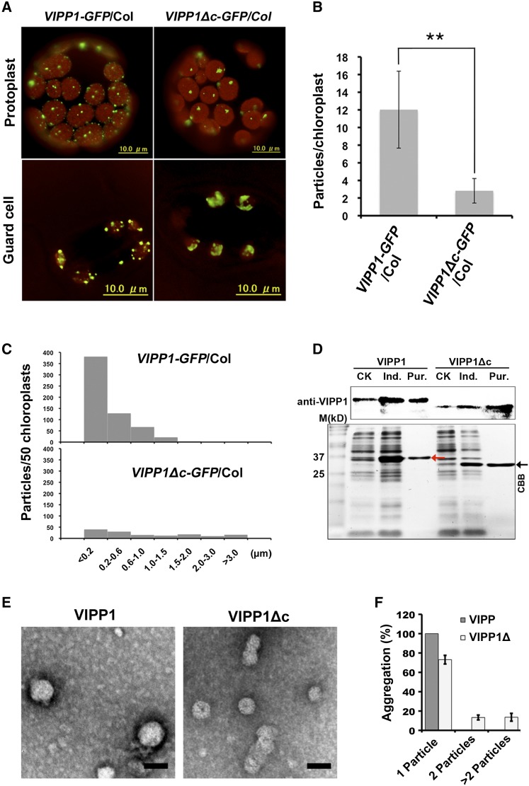
Figure 2.
Effects of Vc on the clustering of VIPP1 particle in Arabidopsis. A, Representative mesophyll protoplasts and guard cells of VIPP1-GFP/Col (left) and VIPP1Δc-GFP/Col (right). Chlorophyll autofluorescence and GFP signals are shown as red and green, respectively. B, Number of GFP particles per chloroplast in protoplasts from VIPP1-GFP/Col (left) and VIPP1Δc-GFP/Col (right; n = 50, **P < 0.01). C, Histograms showing the distribution of different sizes of GFP particles (estimated by the maximal diameter of each particle) are shown (n = 50). D, Expression and purification of His-tagged VIPP1 and VIPP1Δc fusion proteins in E. coli. Red and black arrowheads, respectively, indicate the VIPP1 and VIPP1Δc positions. E, Recombinant VIPP1 and VIPP1Δc expressed and purified from E. coli in P buffer were visualized using negative stain and subsequent observation by TEM. Bars = 40 nm. F, Clustering of VIPP1 and VIPP1Δc particles estimated using the number of self-associating particles (single, two, and more than two particles, n = 30). CK, extracts from bacterial culture without IPTG; Ind., extracts from bacterial culture supplemented with 0.8 mm IPTG for induction; Pur., VIPP1 (left) or VIPP1Δc (right) fusion proteins purified from IPTG-induced E. coli lysates.

