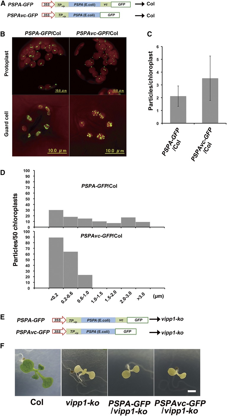
Figure 4.
Effects of Vc on the PspA particle in Arabidopsis and complementation of vipp1-ko mutant with PspA-GFP and PSPAvc-GFP. A, Schematic illustration of PSPA-GFP and PSPAvc-GFP constructs used for transformation into Col. TPvip indicated the transit peptide of Arabidopsis VIPP1. B, Representative mesophyll protoplasts and guard cells of PSPA-GFP/Col (left) and PSPAvc-GFP/Col (right). Chlorophyll autofluorescence and GFP signals are depicted respectively as red and green. C, Numbers of GFP particles per chloroplast in protoplasts of PSPA-GFP/Col (left) and PSPAvc-GFP/Col (right; n = 50). D, Histograms showing the distribution of different sizes of GFP particles (estimated by the maximal diameter of each particle; n = 50). E, Schematic illustration of PSPA-GFP and PSPAvc-GFP constructs used for transforming vipp1-ko. F, Phenotypes of 7-d-old seedlings grown on MS agar medium. Bar = 3 mm.

