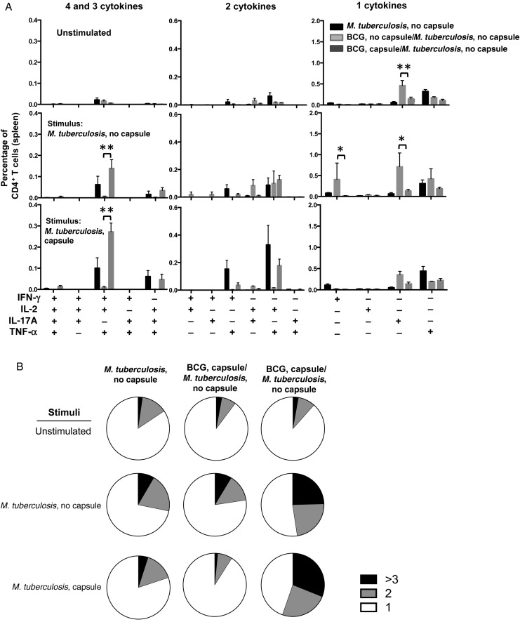
Figure 4.
Analysis of multifunctional CD4+ T cells after challenge with unencapsulated Mycobacterium tuberculosis strain H37Rv. A, Multiparameter flow cytometry with intracellular staining for cytokines in splenocytes from 5 animals immunized subcutaneously 6 weeks previously with 1 × 10⁶ colony-forming units of encapsulated or unencapsulated bacillus Calmette-Guerin (BCG) cells and challenged with an unencapsulated M. tuberculosis strain. Splenocytes were restimulated in vitro with whole-cell lysates from encapsulated or unencapsulated M. tuberculosis plus soluble anti-CD28 monoclonal antibody. The graphs show the percentages of total CD4+ T cells producing interferon γ (IFN-γ), interleukin 2 (IL-2), tumor necrosis factor α (TNF-α), or interleukin 17 (IL-17) and combinations of these cytokines. *P < .05 and ***P < .001, by 1-way analysis of variance with the Tukey post hoc test. B, Pie charts summarizing frequencies of cells producing 1, 2, or 3 cytokines in the experiment shown in panel A. The results are representative of 2 independent experiments.

