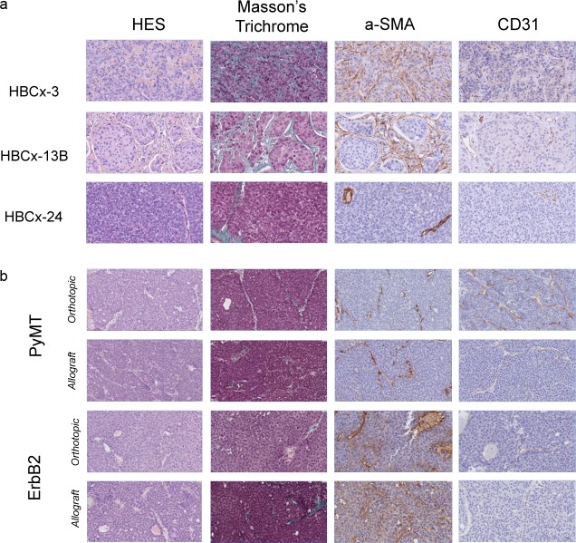
Fig 1. Histology and immunohistochemistry of myofibroblasts and endothelial cells in human and mouse breast cancer tumors.
Slides stained with hematoxylin, eosin and safranin (HES) and Masson’s trichrome, and stained for α-SMA and CD31: (a) human PDXs and (b) murine PyMT and ErbB2 tumors (original magnification: 400x).

