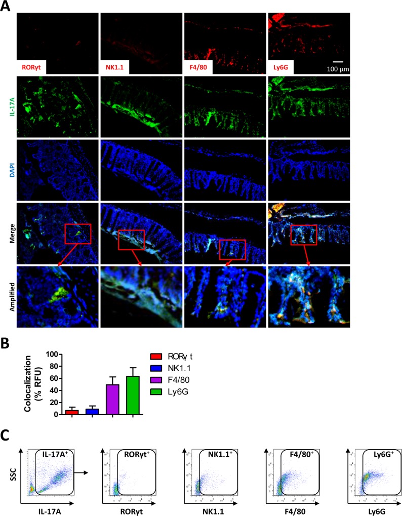
Fig 5. Neutrophils and monocytes are main sources of IL-17A upon CLP.
(A) anxa2 -/- mice were subjected to CLP procedures to induce polymicrobial sepsis. At 24 h post-CLP, colon tissues were sectioned for immunostaining with RORγt, NK1.1, F4/80, Ly6G, and IL-17A antibodies. (B) Colocalization of red fluorescence (to label indicated cell types) with green fluorescence (IL-17A+) quantified as percentage of total green fluorescence. (C) IL-17A positive events in peritoneal lavages from anxa2 -/- mice at 24 h post-CLP. The IL-17A+ cells were collected and further stained with different antibodies and shown as scattergrams. Data are representative from 3 independent experiments. Scale bar = 100 μm.

