
Figure 5.
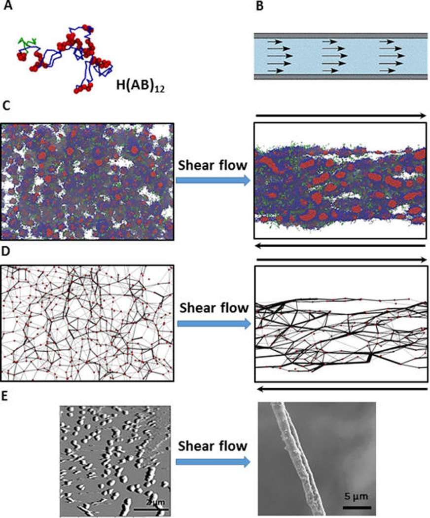
Fiber spinning combining experiment and simulation. (A) Color code of a single recombinant silk protein H(AB)12 in simulation: green, H domain, hexahistidine fusion tag; red, A domain, hydrophobic block; blue, B domain, hydrophilic block. (B) Shear flow in syrange provides flow focusing process of spinning duct of spider. (C) Simulation snapshots after equilibration (left) and after shear flow (right) show that isolated clusters of hydrophobic domains (red spheres) get connected and directed toward shear flow. (D) Post processing of the simulation results. Red circles (nodes) are assigned to the center of the hydrophobic clusters. Two nodes are assumed to be connected by a black line connection (bridge) if part a chain appears in those two nodes. Shear processing increases the connectivity of the network of polymer (E) AFM result on the left shows formation of micels (corresponding to nodes in the simulation). SEM image of the dried fiber on the right shows flow focusing effect after spinning. Reprinted with permission from ref 179. Copyright 2015 Macmillan.