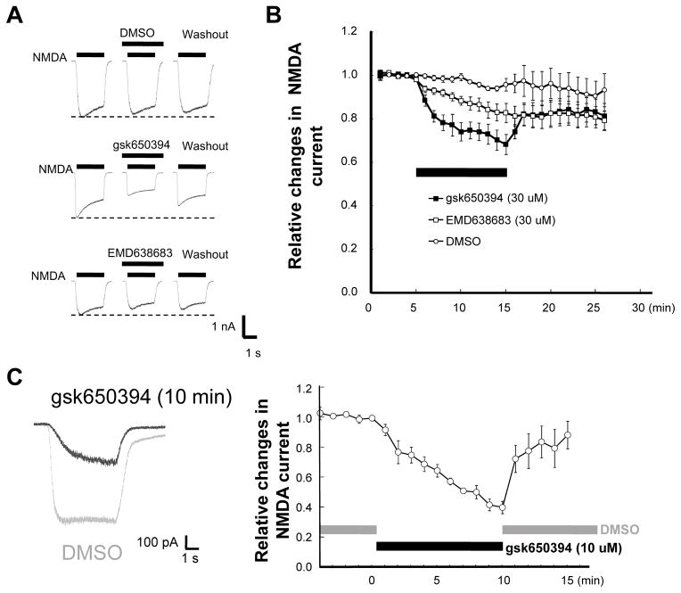
Figure 3. Inhibition of NMDA-induced current by SGK inhibitors.
(A) Representative traces of whole-cell patch-clamp recording showing currents generated by NMDA in cortical neurons in the absence (before) and presence (10 min after treatment) of the indicated inhibitors. (B) Time-dependent changes in normalized peak currents obtained from cells that received different treatments described in (A). (C) Left; representative traces (gray; DMSO, black; 10 μM gsk650394) of gramicidin-perforated patch-clamp recording showing currents activated by NMDA in cortical neurons in the absence (gray, before treatment) and presence of SGK inhibitor (black, 10 min after treatment). Right; time-dependent changes in normalized peak currents obtained from cells with gramicidin-perforated patch-clamp recordings.

