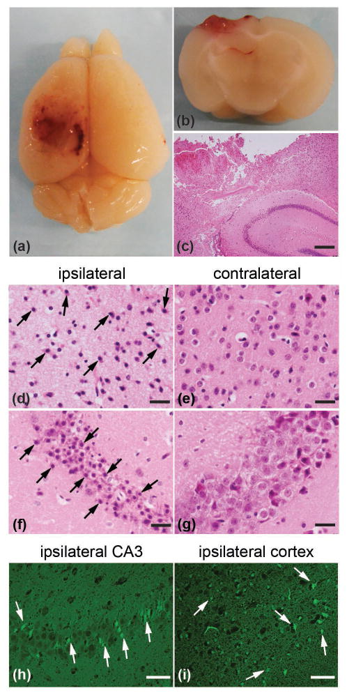
Figure 1.
CCI induces neuropathological changes in the mouse brain. (a) Top view of the mouse brain showing extensive cortical lesion in the left, ipsilateral hemisphere one day after injury. (b and c) Coronal section across area of cortical lesion characterized by loss of tissue, hemorrhage and necrosis in the cortex, as well as subcortical white matter and hippocampus. H&E staining was used in (c). (d and e) Cortical edema and shrunken, pyknotic neurons (arrows indicate characteristic examples) were observed in the injured ipsilateral cortex (d), but not contralateral cortex (e) using H&E staining. (f and g) Loss of pyramidal neurons and shrunken, pyknotic neurons (arrows indicate characteristic examples) were found in ipsilateral hippocampal area CA3 and CA2 (f), but not contralateral CA3 and CA2 (g) using H&E staining. Degenerative neurons (arrows indicate characteristic examples) in ipsilateral hippocampal area CA3 and cortex were confirmed by Fluoro-Jade B (FJB) staining (h and i). Scale bars: 200 μm (c); 50 μm (d, e, h and i); 20 μm (f and g).

