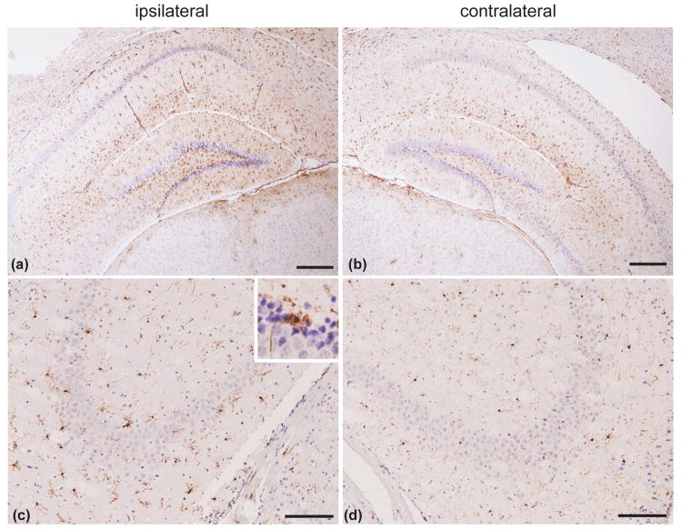
Figure 2.
CCI induces neuroinflammation in the mouse brain. (a and b) Neuroinflammation indicated by increased astrocyte activation in the ipsilateral hippocampus (a), as compared to the contralateral hippocampus (b) using GFAP immunostaining. (c and d) Neuroinflammation indicated by increased number of reactive microglia in the ipsilateral hippocampal area CA3 and CA2 (c), as compared to contralateral CA3 and CA2 (d) using Iba1 immunostaining. Insert in panel (c), represents magnification of a region in hippocampal area CA3 with activated microglia. Scale bars: 200 μm (a and b); 100 μm (c and d).

