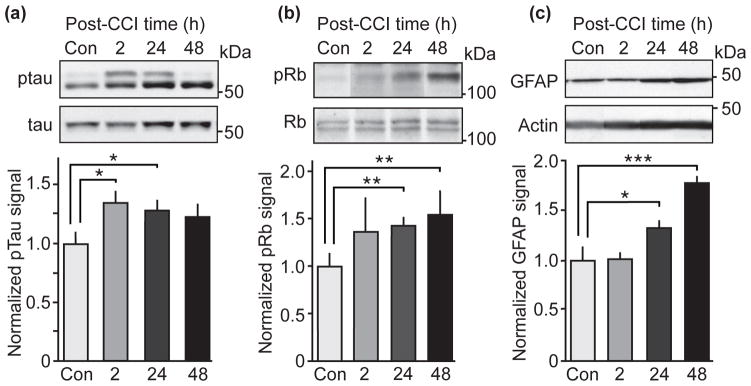
Figure 4.
Analysis of Cdk5-dependent phosphorylation of tau and Rb protein and GFAP levels in hippocampal lysates at various post-CCI time points. (a, b and c) Quantitative immunoblot analysis from lysates of ipsilateral hippocampus at 2, 24 and 48 hours post-CCI probed for tau phosphorylation at Ser202 (CP13; Fig. a), Rb protein phosphorylation Ser807/811 (b) and GFAP levels (c). Representative immunoblots and corresponding quantifications are shown. Molecular weights are indicated in kDa on the right side of the immunoblots. All data are presented as mean ± SEM; n = 4–5; *p < 0.05, **p < 0.01, ***p < 0.001; Student’s t-test.

