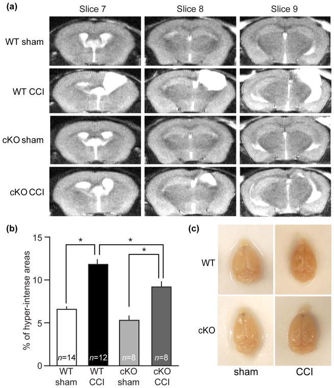
Figure 5.
Loss of Cdk5 attenuates CCI-induced lesion volume. (a) Representative MRI images of axial slices from conditional Cdk5 knockout mice (Cdk5 cKO) and wildtype littermates (WT), acquired at 28 days post-CCI. (b) Hyper-intense areas were expressed as percentage of total brain volume. (c) Top view of dissected brains from Cdk5 cKO and WT showing reduced lesion in Cdk5 cKO. All data are presented as mean ± SEM; WT sham n = 14, WT CCI n = 12, cKO sham n = 8 and cKO CCI n = 8; *p < 0.05, One-way ANOVA with Turkey’s multiple comparison test.

