Figure 5.
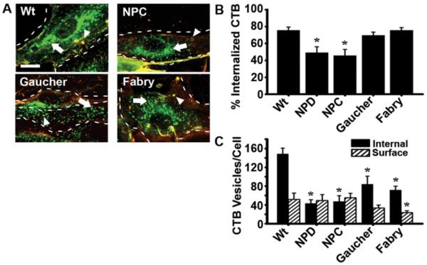
Internalization of cholera toxin B in LSD fibroblasts. Wild-type (Wt), NPD, NPC, Gaucher, and Fabry fibroblasts incubated with fluorescent cholera toxin B (CTB; green pseudocolor) for 1 h at 37°C. (A) Fluorescence images were taken after unbound CTB was washed off and surface-bound CTB was immunostained (red pseudocolor). This rendered surface-bound CTB marked in two colors (red + green = yellow; arrowheads), distinguishable from internalized CTB (green; arrows). NPD, reported in 29 serves as a control and is not shown. Dashed lines = cell borders, as observed from phase-contrast microscopy. Scale bar = 10 μm. (B) Quantification of the percentage of single-labeled CTB found inside each cell compared to total CTB (single-labeled and double-labeled) associated with said cells. (C) Internal and surface CTB (fluorescent objects over background). (B,C) Data are the mean ± SEM. *Comparison with Wt (p<0.05 by Student’s t-test).

