Fig. 1.

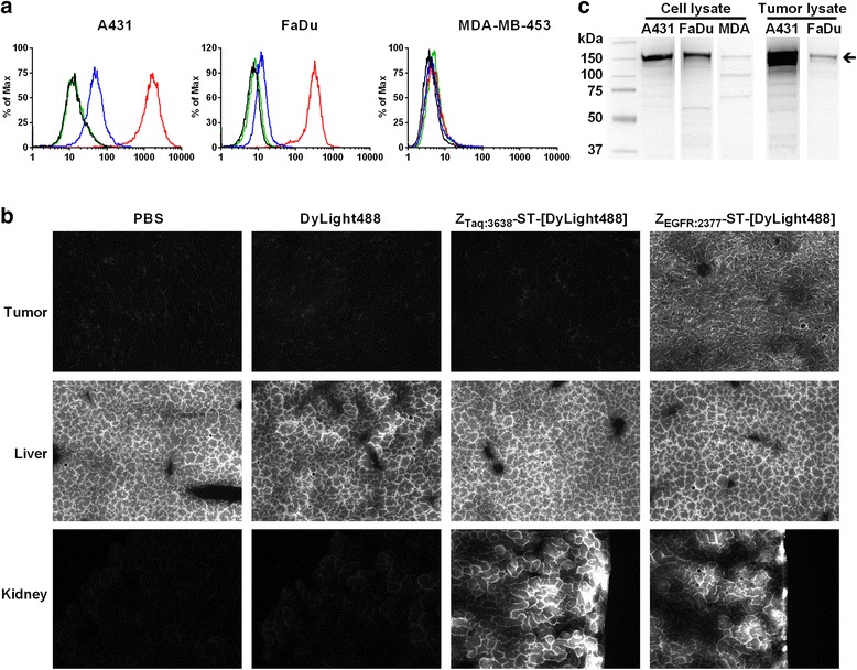
a Cell-binding assay of non-blocked (red) and blocked (blue, tenfold ZEGFR:2377) ZEGFR:2377-ST-[DyLight 488] and controls ZTaq:3638-ST-[DyLight 488] (green) and DyLight 488 dye (black) using FACS analysis in A431 (left), FaDu (middle), and MDA-MB-453 (right) cells with high, medium, and low/no expressions of EGFR, respectively. b Fluorescent microscopy images of tumor (A431), liver, and kidney. Fluorescence in the tumor is only observed with the ZEGFR:2377 probe. High autofluorescence of the liver is observed with all probes. Fluorescence from both the ZEGFR:2377 and ZTaq:3638 probes is observed in the kidney. c western blots of A431, FaDu, and MDA from cell (left) and tumor (right) lysates using an antibody against human EGFR. The protein concentration of the cell lysates was determined by Bradford protein assay and Ponceau S staining of the membranes was used as loading control. The arrow indicates full-length EGFR
