Fig. 4.

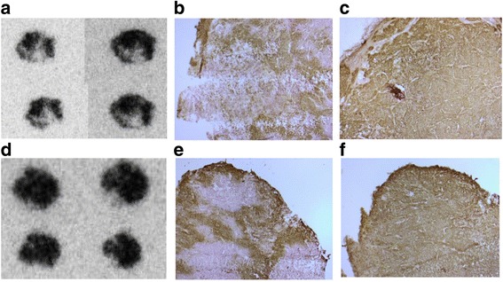
Ex vivo analyses of sections of the two tumors in Fig. 3b (left tumor (a–c); right tumor (d–f)). a, d Phosphoimaging of sections of tumors excised immediately after the 60 min PET scan with [methyl-11C]-ZEGFR:2377-ST-CH3. b, e IHC detecting CAIX, staining was performed on formalin-fixed, paraffin-embedded material using antibody against CAIX (BioScience, Slovakia) at a dilution 1:500. c, f IHC detecting EGFR, staining was performed on formalin-fixed, paraffin-embedded material using antibody against EGFR (Sigma, Sweden) at a dilution 1:800
