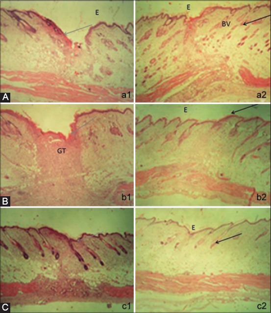
Figure-4.

The histology of skin tissues at the 3rd days of human umbilical mesenchymal stem cells conditioned medium (HU-MSCM) treatment stained with hematoxylin and eosin (H and E). (a2) Small number of inflammatory cells and abundance of blood vessels (black arrow) in compared with the control which still have abundance inflammatory cells (gray arrow) (a1) (magnification 20×) E: Epidermis, BV: Blood vessels. (b) The histology of skin tissues at the 6th days of human umbilical mesenchymal stem cells conditioned medium treatment stained with hematoxylin and eosin (b2), Decreasing inflammatory cells followed by increasing re-epithelization and more density of collagen fiber (black arrow), (b1) sample from the control, a lot of inflammatory cells are found (gray arrow) (magnification 20×) E: Epidermis, GT: Granulation tissues. (c) The histology of skin tissues at the 9th day of human umbilical mesenchymal stem cells conditioned medium treatment stained with hematoxylin and eosin. Fibroblasts are increasing and hair follicles created, the muscles regenerations is clearly appeared (black arrow) (c2), however, on the povidone-iodine treatment, infiltration of inflammation cells are still increasing (gray arrow) (c1) (magnification 20×). E: Epidermis.
